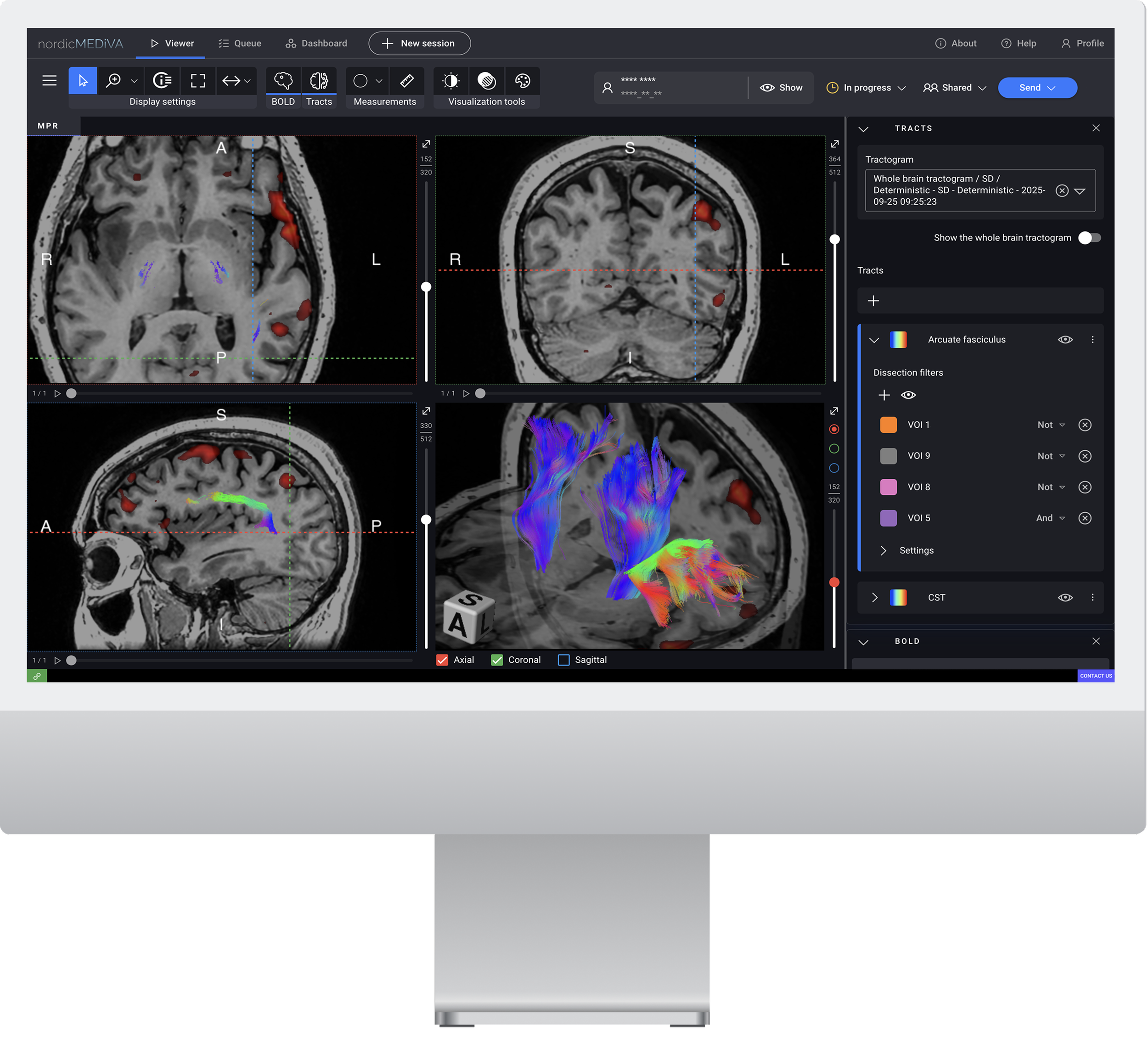
Next-generation neuroimaging software from NordicNeuroLab
Discover automatic processing and powerful visualization backed by
20 years of experience in the clinical fMRI market (nordicICE and nordicBrainEx)
Web-based viewer
〰️
Automatic processing
〰️
Live collaboration
〰️
Cloud-based installation
〰️
Fast responsiveness
〰️
Customizable interface
〰️
Flexible ROI analysis
〰️
Flexible exporting options
〰️
Web-based viewer 〰️ Automatic processing 〰️ Live collaboration 〰️ Cloud-based installation 〰️ Fast responsiveness 〰️ Customizable interface 〰️ Flexible ROI analysis 〰️ Flexible exporting options 〰️
Modules
-

Task-based fMRI
Automated BOLD fMRI pre-processing pipeline which aligns with the QIBA guidelines. Processing includes 3D spatial smoothing, and statistical analysis using the General Linear Model.
-

Diffusion and Tractography
Advanced diffusion analysis with both standard DTI and SD algorithms to better resolve areas with crossing fibers and a more complete anatomical representation. Over 10 diffusion maps for in-depth insights. Improved image quality with Eddy current and motion correction.
-

DSC Perfusion
Fully automated T2* perfusion processing. Choice of leakage correction algorithms. Automatic export to PACS.
Trusted by top-ranked hospitals
Contact us to get a free trial
nordicMEDiVA is available for sale only in the United States.
Get login credentials within 24 hours
Integration and IT department involvement are not required
Access trial on our servers from a web browser
Use our data or send us your data anonymized
No paperwork!
Streamline your workflow
Experience the benefits of our fast, web-based, vendor-neutral viewer and auto-triggered processing. Automate large workflow parts, saving time for the activities that matter.
Collaborate in real-time
Our live collaboration lets you share sessions with ROIs and data synced in real time. Ask a colleague for a second opinion or interactively discuss a case with the neurosurgeon.
Integrate easily and securely
Choose between on-premise installation or our cloud-based solution for safe and convenient integration.
Standardize data processing
Define the automated image analysis pipelines and settings at the organization level. Set up multiple routes tailored to specific clinical cases and control access permissions for added security.
Control the data quality
Automatic motion correction and coregistration ensure confidence in your results. nordicMEDiVA provides you with tools for quality control, such as motion correction graphs and fMRI intensity curves overlayed on a paradigm. Coregistration is triggered when loading maps and performed automatically with the option for manual correction.
Export to neuronavigation and PACS
With just a few clicks, export all directional slice stacks to PACS as secondary captures, including associated data such as region of interest (ROI) statistics and intensity curves. Send merged overlays on structural datasets to neuronavigation systems or export any raw maps for further analysis.
Tailored solutions for your clinical needs
The nordicMEDiVA modules are FDA-cleared (K243209) and available for sale in the Unites States.
Pre-surgical planning with fMRI and Tractography
-
Automatically generate activation maps from BOLD fMRI time series.
Produce statistical maps using the general linear model, motion correction, spatial and temporal smoothing, and automatic noise thresholding (Otsu).
Adjust the threshold interactively in the viewer.
-
Access both Diffusion Tensor Imaging (DTI) and the improved Spherical Deconvolution model, gaining flexibility to select the optimal approach for every clinical scenario.
Dissect tracts using an ROI-based approach.Output common maps like FA, MD, and more to quantify the diffusion in areas of interest.
Export results as both a 3D tract object and fused with the anatomical scan, which is compatible with most neuronavigation systems.
Tumor evaluation with
fully-automatic DSC
Benefit from advanced T2* perfusion processing with automatic global AIF search and fast calculation of perfusion maps such as CBV and CBF.
Choose between standard leakage correction based on the Weisskoff method, or using a residue-function based approach that’s insensitive to changes in MTT.
Maps are automatically normalized to normal-appearing tissue and can provide quantitative data by comparing ROI values from different anatomical regions.
Analyze dynamic time curves for comprehensive evaluation of perfusion data.
Automatically output maps directly to PACS, offering significant potential for cost savings and efficiency.
User interface features
-
Multi-modality support
Analyze different modalities in one session. The relevant tools automatically launch based on a loaded map, streamlining your workflow.
-
Customizable layout
Arrange and resize analytical tools (widgets), maximize individual MPR planes, or showcase your work in full-screen mode for presentations, with the view saved for the next time you open this session.
-
Visibility control
Toggle on and off the visibility of maps, Protected Health Information (PHI), ROIs, distance measurements, MPR headsup information, graphs, and statistical values to tailor the display to your preferences.
-
Multi-patient management
Keep multiple patient sessions open simultaneously across different browser tabs.
-
Shared access between sites
Share a single software installation across multiple hospitals or imaging centers of your organization for easy collaboration and consistent analysis.
-
Flexible map loading
Easily add series to the MPR with drag-and-drop functionality, and load as many maps as needed for comprehensive analysis.
Don't just trust our word
FAQ
-
We support BOLD fMRI, DWI, and DSC acquisitions from all major scanner vendors — including data from 7T scanners. Our platform also handles multi-shell DWI for tractography and supports both enhanced and standard DICOM formats.
-
Yes! DICOM-compatible and capable of analyzing data from all major MRI vendors.
-
Yes! The BOLD fMRI, tractography, and DSC perfusion modules can be purchased independently.
-
Yes! An online trial can be arranged with your anonymized data. Please contact your local sales representative or sales@nordicneurolab.com for more details
-
An online trial can be arranged, either with our demonstration data or with your own data. The demo is accessed on the internet — no local installations or involvement from local IT departments is needed.
-
-
-
We offer hands-on training for post-processing and analyzing collected MRI data using the nordicMEDiVA software. For task-based fMRI, this includes applying design files, verifying data accuracy, and creating and interpreting activation maps. If you choose to use other modules of nordicMEDiVA, such as Diffusion and Tractography or Perfusion MRI, we provide comprehensive training on those as well. You will learn how to summarize your findings and export data in the appropriate formats.
Regardless of your post-processing pipeline, we can advise on general pre-processing and analysis steps to support your work.
-
For the US market, our main office is located in Milwaukee, WI and we have a local presence with sales and application specialists in West, South, and East regions.
-
Yes. We are compatible with DICOM to ensure compatibility with MR vendors, PACS systems, and neuronavigation systems.


